Welcome to the Digital Age of Dentistry
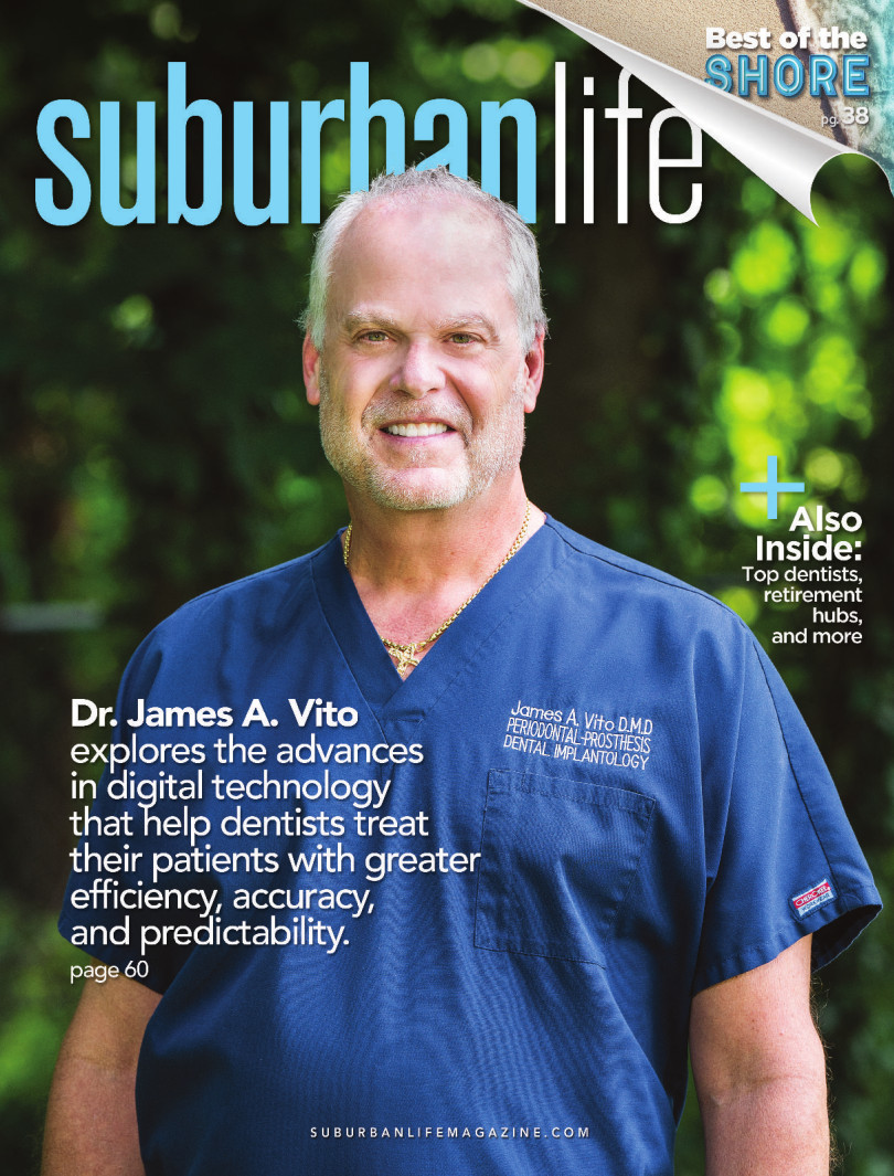
Dr. James A. Vito explores the advances in digital technology that help dentists treat their patients with greater efficiency, accuracy, and predictability.
The digital age of dentistry has arrived, and numerous benefits have come with it: a higher quality of dental care, more reliable and accurate information, and a better overall experience for patients. Because of these advances, patients can now expect faster results and an enhanced ability to identify dental issues before they happen.
Digital platforms represent the future of dentistry. In fact, they are already changing the way we provide dental care. Digital radiographs, which have become the norm for most dental practices, allow us to gather and store more accurate data on patients than ever before.
CAD/CAM-created restorations are proving to meet and even exceed our expectations of fit, accuracy, and aesthetics. Computerized tomography scan technology (CBCT) helps us plan and identify dental issues with accuracy never before thought possible, and have proven invaluable for the placement and management of dental implants.
Our charts are paperless and contain various types of digital data, such as digital X-rays, CBCT scans, digital restorative scans, intraoral photos, videos, and models of teeth. We use email and texting to notify patients of appointments and other news pertaining to our practice.
Digital X-rays are safer and more effective than the traditional alternative. The sensor produces 90 percent less radiation than traditional X-ray film, and also creates clearer images for dentists and their office staff. These images can be transferred directly to a computer and then magnified, enhanced, and rotated for better analysis. In other words, as the following nine bullet-pointed items illustrate, we are able to get a clearer idea of a patient’s bite structure, how well the jaws align, and the underlying health of the mouth.
* Enhanced images: Digital sensors produce high-resolution images for use by the dentist and his or her staff. In addition to images of the teeth, these X-rays can capture images of the gums and other oral structures.
* Enlargement options: When you are looking at a traditional X-ray film, the image can be viewed only at actual size. Digital images can be enlarged, giving the dentist the option to zoom in on potential problems.
* Immediate viewing: Traditional X-ray film required a waiting period while the film was developed. Now, the digital tools provide immediate viewing of the imaged area.
* Improved diagnostics: Having access to clear pictures and more information increases the accuracy of diagnostics, so a diagnosis and treatment plan can be customized to the exact specifications of a patient’s mouth. Digital X-rays can reveal inconspicuous issues such as hidden areas of decay, infections in the bone, gum disease, tumors, and other abnormalities that may not be apparent during a visual examination.
* Savings of time and money: Early detection methods using digital X-rays help to minimize required treatments. As a result, patients can save both time and money by avoiding more invasive treatments.
* Reduced radiation: Researchers have found that radiation levels are reduced by up to 70 percent compared to traditional X-ray equipment. Lowering the radiation helps to decrease the potential side effects and long-term risk of X-ray exposure.
* Electronic storage: The dental office can keep these files in storage indefinitely. Instead of having stacks of paperwork and files, digital images can be kept securely in the cloud for future reference as needed.
* Easy access: Digital images can be sent through electronic systems to insurance companies, helping to speed up the process for certain insurance claims. They can also be printed and saved for easy storage.
* Environmentally friendly: Digital X-rays reduce pollution because they are produced without the hazardous chemicals needed to produce traditional X-rays.
Digital Advances for Dental Implants, Endodontics, and More
The benefits of digital technology extend to CBCT scans, which provide more information than a conventional dental X-ray, allowing for more precise treatment planning. CT scanning is painless, noninvasive, and accurate. A major advantage of CT is its ability to image bone and soft tissue all at the same time, and provide a 3D rendering of the area(s) in question. No radiation remains in a patient’s body after a CT exam.
The benefits of digital technology extend to CBCT scans, which provide more information than a conventional dental X-ray, allowing for more precise treatment planning. CT scanning is painless, noninvasive, and accurate. A major advantage of CT is its ability to image bone and soft tissue all at the same time, and provide a 3D rendering of the area(s) in question. No radiation remains in a patient’s body after a CT exam.
Another exciting and growing aspects of digital dentistry is digital intraoral impressioning. It is now possible to capture accurately impression data more quickly than when using conventional impression materials. Digital impressions eliminate the need for messy impression materials that remain in a patient’s mouth for several minutes, while providing a more comfortable experience for patients, especially those who have a strong gag reflex.
When done correctly, digital impressions can increase productivity, efficiency, and accuracy. Turnaround times may be faster because the digital file is instantly sent to the dental laboratory to create the restoration, with no need to cast up a traditional working model. These scans provide the same data in a cleaner, more accurate fashion, and they also give the dentist a permanent model of teeth, which then can be stored indefinitely by both the dentist and dental lab. Digital impressions are also eco-friendly, because the process eliminates the need for disposable impression trays and materials that would otherwise end up in a landfill.
A digital scan can be magnified and evaluated by the dentist while the patient is still in the chair; if there are any errors, they can be corrected before the file is submitted to the dental lab. The ability to see the impression on-screen allows the dentists to check the tooth preparations for accuracy and allows for modification of the tooth preps, if required, at the same appointment. This saves the patient from having to come back for a new impression.
CBCT scans allow for the precise placement of dental implants in the bone, proper orientation of the implant with its overlying restoration, prevention of injury to nerves, prevention of implant penetration into the sinus, and selection of the right-sized implant for optimal support. Also, the evaluation can determine whether bone grafts are needed to improve the height and/or width of bone. If bone grafts have been performed, these scans can determine whether they have worked, as well as the evaluation of the density of the bone. CBCT scans also allow for restorative planning with the dental lab prior to implant placement, and allow for the coordination of both the implant placement and resulting restoration for the optimal functional and aesthetic result.
In 2015, the American Academy of Endodontists and the American Academy of Oral and Maxillofacial Radiology issued a revised joint position statement on the use of CBCT in endodontics. The joint statement indicates the effectiveness of 3D imaging for endodontic diagnosis and treatment because it demonstrates anatomic features in 3D that intraoral, panoramic, and cephalometric images cannot.
For endodontic therapy, CBCT scans are more accurate than conventional X-rays or Panorex images. CBCT scans allow the endodontist to see the tooth anatomy, root forms, and number of canals of the tooth in question, as well as the size and scope of the pathology, the detection of root fractures, root resorption, and postoperative assessment.
In conclusion, dentistry has caught up with most dental issues. As a result, patients can expect better dental care, making simple and complicated dental treatment more efficient, predictable, and accurate than ever.
James A. Vito, D.M.D.
523 E. Lancaster Ave.
Wayne, PA 19087
(610) 971-2590
www.jamesvito.com
523 E. Lancaster Ave.
Wayne, PA 19087
(610) 971-2590
www.jamesvito.com
Photo by Jeff Anderson
Published (and copyrighted) in Suburban Life magazine, July 2022.


